
Figure 6.
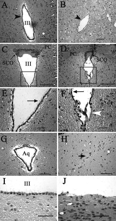
Alterations in the ventricular system of hydrocephalic Ro1 mice. Hematoxylin and eosin staining of coronal sections from single-transgenic control (P45, left column) and Ro1 (P33, right column) mice. A, B, In Ro1 mice (B), the third ventricle at the level of the periventricular hypothalamic nucleus is reduced in size and the ependymal cell layer appears thinner compared with controls (A). D, F, H, J, The subcommissural organ is disorganized in Ro1 mice (D), and there is a partial denudation of ependymal cells lining the ventricular walls (F, arrowhead; J). The remaining ependymal cells have fewer cilia (F, arrow), and the aqueduct is obliterated with no apparent ependymal cell layer (H). C, E, G, I, In control mice, the subcommissural organ (C) and aqueduct (G) show normal morphology. The ependymal layer is intact with multiple cilia per cell (E, arrow; I). E and F are enlargements of the boxes in C and D, respectively. Mice were maintained off dox. Aq, Aqueduct; SCO, subcommissural organ; III, third ventricle; PC, posterior commissure. Scale bars: A, B, G, H, I, J, 50 μm; C, D, 100 μm; E, F, 30 μm.