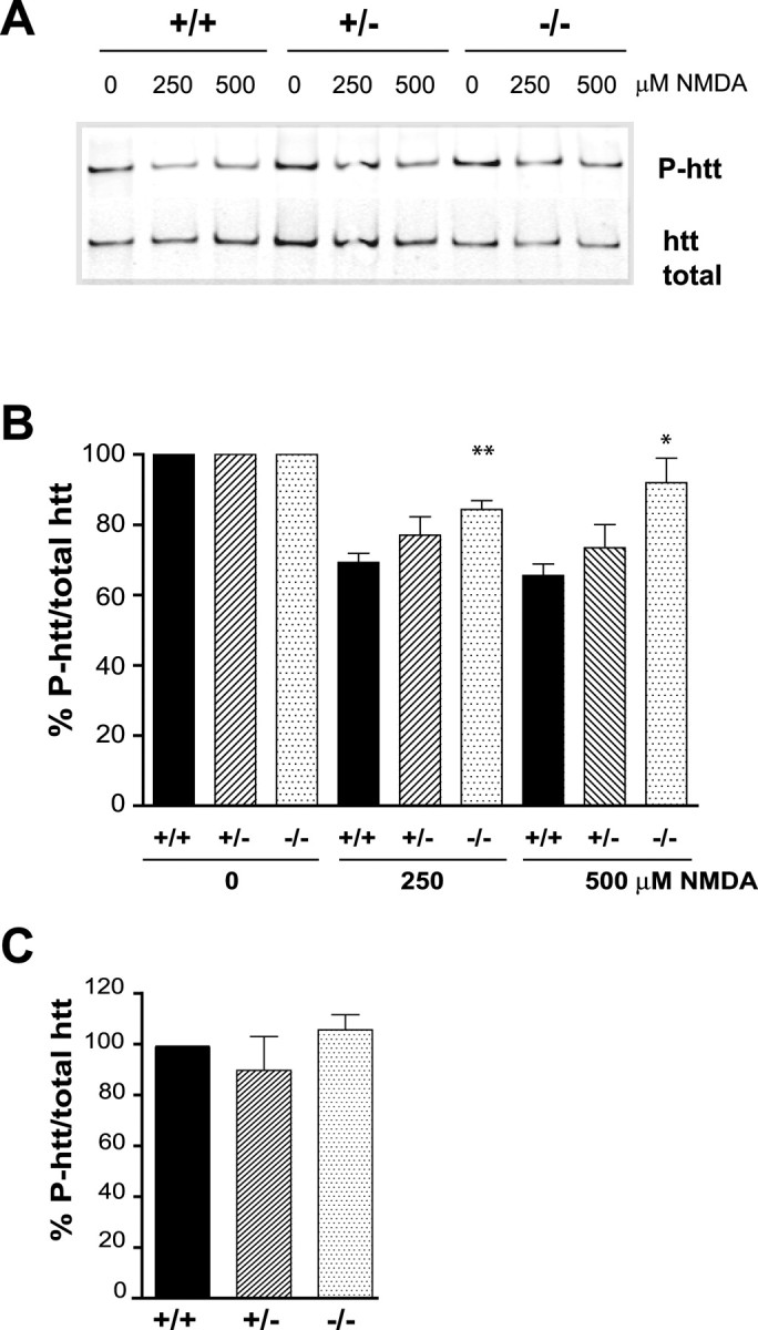
Figure 5.

HIP1 promotes dephosphorylation of htt during NMDA-induced excitotoxicity. A, Cortical neurons from wild-type and HIP1−/− mice were treated with varying doses of NMDA and 30 μm glycine for 10 min at 12 DIV. After drug removal, neurons were cultured for 90 min and processed for immunoprecipitation of htt. Precipitated protein was separated by Western blot, and membranes were probed with Abs against P-htt-S421 and total htt (mAb 2166). B, The ratio between P-htt-S421 and total htt was determined by densitometry and expressed relative to controls. Ratios for each condition and genotype in each of six independent experiments were determined (*p < 0.05; **p < 0.005 compared with wild type; t test). C, The absolute ratio between P-htt-S421 and total htt was determined by densitometry under control conditions for all three genotypes and expressed relative to wild type (n = 4). Error bars denote ±SEM.
