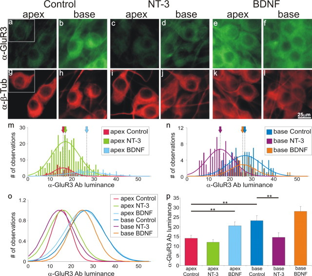
Figure 4.
BDNF increases, whereas NT-3 reduces, anti-GluR3 antibody labeling in spiral ganglion neurons. Spiral ganglion neurons were double labeled with anti-GluR3 antibody (a–f; green) and anti-β-tubulin (α-β-Tub) antibody (g–l; red). Neuronal labeling was assessed under control conditions (no added neurotrophin; a,b and g,h), exposure to media supplemented with 5 ng/ml NT-3 (c,d, and i,j), or media supplemented with 5 ng/ml BDNF (e,f and k,l). m, Frequency histograms and Gaussian fits to measurements made from apical neurons in control media (red) compared with neurons in NT-3-supplemented (green) and BDNF-supplemented (light blue) media from a single experiment. Despite the differences in peak amplitude, the mean luminance (dotted line/arrow) is clearly shifted to the right when BDNF is added to the culture medium. n, Frequency histograms and Gaussian fits to measurements made from basal neurons in control media (blue) compared with neurons in NT-3-supplemented (purple) and BDNF-supplemented (orange) media from a single experiment. The mean luminance (dotted line) is shifted to the left when NT-3 is added to the culture medium. o, Normalized Gaussian fits for histograms shown in m and n. p, An average of seven separate experiments shows that the differences in anti-GluR3 antibody labeling in the presence of BDNF and NT-3 are statistically significant. Scale bar in l applies to a–l.

