
Figure 1.
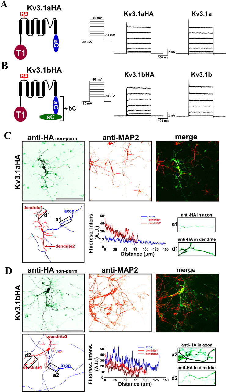
Distinct targeting patterns of Kv3.1 splice variants. HA-tagged Kv3.1a (A) and Kv3.1b (B) channels are functional and similar to the wild type in activation potential (approximately −10 mV) and current amplitude. Structural diagrams are on the left. Transmembrane segments are shown with six black bars. The HA tag (in red) is within the first extracellular loop. The T1 domain (dark red), the C-terminal region of Kv3.1a (in blue), and the splice domain in Kv3.1b (in green) are indicated. HA-tagged and wild-type channels were subcloned in pIRES2EGFP vector, expressed in HEK293 cells, and recorded by whole-cell voltage clamp. The cells expressing channel constructs were identified with GFP fluorescence, held at −80 mV, and given 250 ms voltage episodes from −50 to 40 mV with a 10 mV increment. Examples of current traces from HA-tagged and wild-type channels are given in middle and right panels, respectively. The endogenous voltage-gated outward currents only reached ∼100 pA at 40 mV and are therefore ignored (data not shown). Shown are representatives of 6–10 cells. C, When expressed in cultured hippocampal neurons, Kv3.1aHA was predominantly localized to the somatodendritic membrane. Neurons were transfected with Kv3.1aHA at 6 DIV, fixed, and stained with an anti-HA antibody (in green) under nonpermeabilized conditions 2 d later. Dendrites of the neurons were labeled by anti-MAP2 (microtubule associated protein 2) staining (in red). The camera lucida drawing of the neuron in the bottom left shows dendrites in red and axons in blue. The anti-HA fluorescence profiles along the major axon (within 200 μm from the soma) and two dendrites (including the entire dendrite, indicated by thicker lines in the drawing), and two boxed areas (in 3 times higher magnification) are shown in the bottom middle and right, respectively. Boxed areas were chosen along the major axon/dendrite processes ∼50 μm away from the soma. D, Kv3.1bHA was preferentially localized to the axonal membrane. Distance 0, Soma. Scale bars, 100 μm.