
Figure 7.
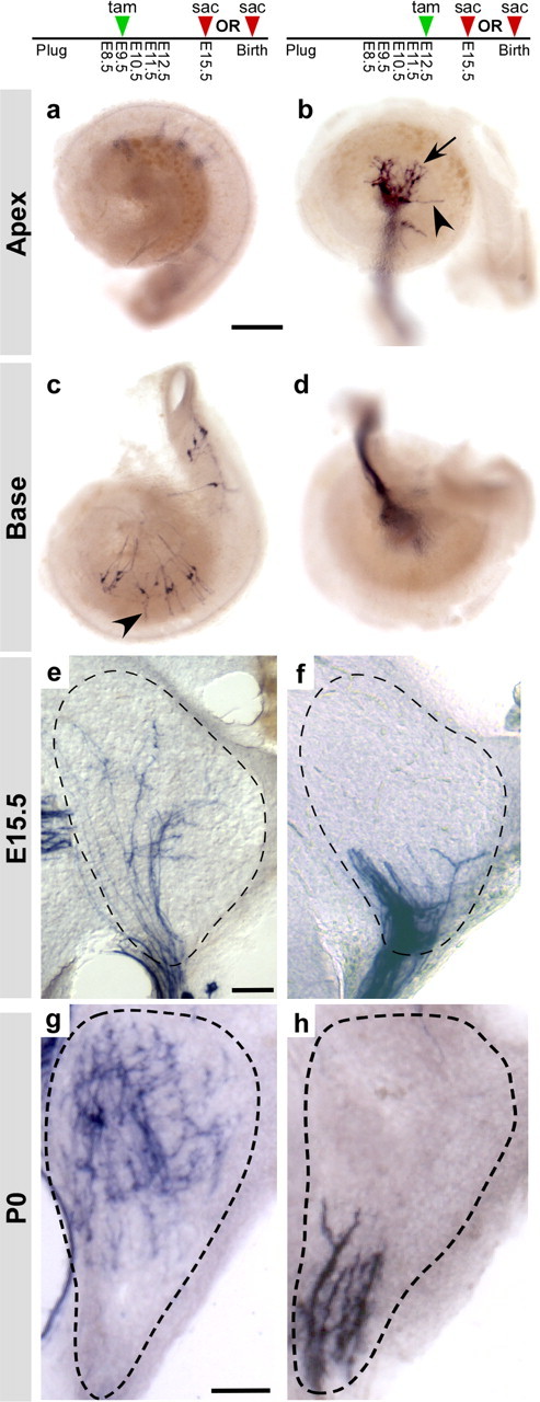
Early tonotopic organization of auditory circuits. a–d, As seen at P0, early tamoxifen (tam) treatments labeled E15.5 neurons in the base but not in the apex of the cochlea (a, c), whereas later treatments labeled neurons in the apex but not in the base (b, d). Cochlear ganglion neurons are at an early stage of circuit assembly, particularly in the apex where processes formed a tangled network close to the cell bodies (arrow, b), with only occasional extensions (arrowheads, b, c) toward the region where hair cells will eventually develop. Ngn1-CreERT2; Z/AP animals were treated with 0.5 mg of tamoxifen on E9.5 (a, c) or on E12.5 (b, d). Cochleas were collected (sac) at E15.5 and stained for PLAP activity. e–h, Although the cochlear nucleus anlage is not cellularly or morphologically mature at E15.5, cochlear ganglion projections were tonotopically organized, with endings from high-frequency neurons restricted to the dorsal half of the nuclei (e) and endings from low-frequency neurons located ventrally (f). This tonotopic organization was similar to what was observed at P0 (g, h). Ngn1-CreERT2; Z/AP animals were given 0.5 mg of tamoxifen on E9.5 (e, g) or E12.5 (f, h) and collected either at E15.5 (e, f) or P0 (g, h). Brains were sectioned coronally; the cochlear nuclei are outlined with dashed lines. For P0, only the posterior ventral cochlear nucleus (PVCN) is shown. Scale bars: a, 250 μm; e, g, 50 μm.