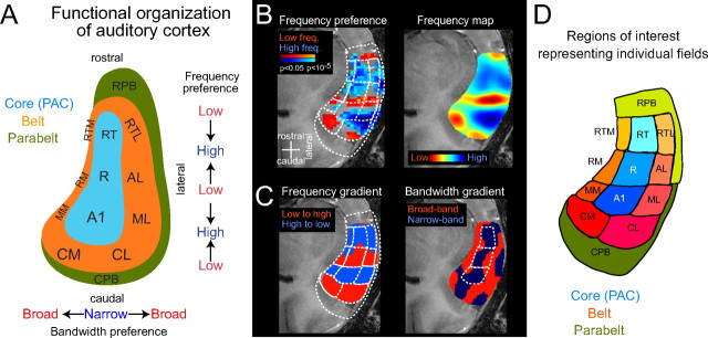
Figure 2.
Functional organization of auditory cortex: localizing auditory cortical fields. A, Schematic of auditory cortex, which can be separated into core (primary auditory cortex), belt, and parabelt regions, adapted from Hackett et al. (1998a). The preference for sound frequencies changes in the rostrocaudal direction and shows multiple reversals. In the orthogonal direction, the preference for sound bandwidth changes from core to belt. Abbreviations of the auditory cortical fields are shown (see below). B–D, The differential preferences for sound frequency and bandwidth can be used to obtain a functional parcellation of numerous auditory fields (for details, see Materials and Methods). Borders were delineated between regions with opposing frequency gradients at the point of mirror reversal and between regions selective to narrow or broadband sounds. B, First, the main frequency-selective regions are approximated by using low- and high-frequency sounds (500 Hz and 8 kHz), as shown by the activation map on the left (p values are color-coded on an anatomical image). Then, a more detailed frequency preference map was obtained by using multiple frequency bands (6 frequencies, equally spaced between 250 Hz and 16 kHz), as shown on the right. C, A gradient analysis (computed along the rostrocaudal direction) results in an alternating pattern of regions with progressively increasing or decreasing frequency selectivity and separates the regions with mirror reversed-frequency gradients. Based on the gradient analysis, borders can be delineated from the points at which the sign of the frequency gradient changes. The same gradient analysis is done for sound bandwidth (tone vs noise preference maps) along the mediolateral direction (right image). D, A functional parcellation of auditory cortex into 11 core and belt fields is obtained by combining bandwidth and frequency maps. As additional ROIs, the caudal and rostral parabelt (CPB and RPB) were defined; these extend rostrocaudally on the superior temporal plane and laterally on the superior temporal gyrus. RT, Rostrotemporal; AL, anterolateral; AM, anteromedial; RM, rostromedial; RTL, rostrotemporal-lateral; RTM, rostrotemporal-medial field.

