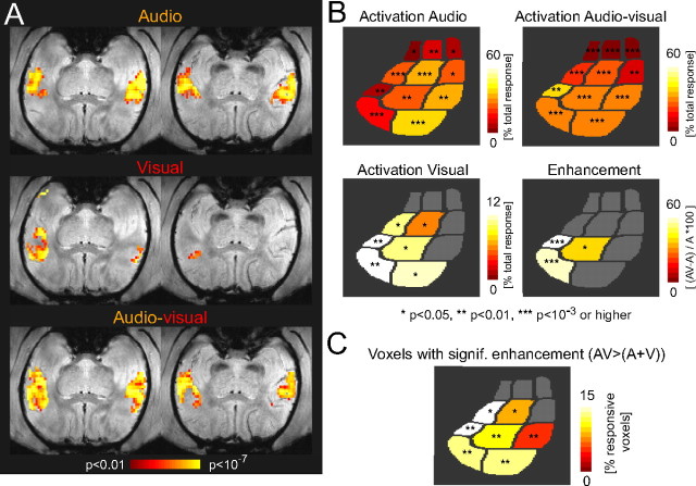
Figure 4.
Activations and multisensory enhancement in the alert animal. A, Example data (two slices covering auditory cortex) from one session with the alert animal. Individual panels display the activation maps for each condition (p values) superimposed on an anatomical image. B, Schematic of core and belt fields showing the activation strength in individual conditions for all experiments with the alert animal (n = 7). Activations are quantified as the relative response (Fig. 3). The bottom right panel displays the percentage enhancement of signal change from auditory to audiovisual conditions. C, Fraction of responsive voxels within individual fields that showed significant nonlinear enhancement. In all panels, color-coding indicates the mean across experiments, and only fields with significant activation or enhancement are colored: t test across experiments; *p < 0.05; **p < 0.01; ***p < 0.001 or higher.

