
Figure 5.
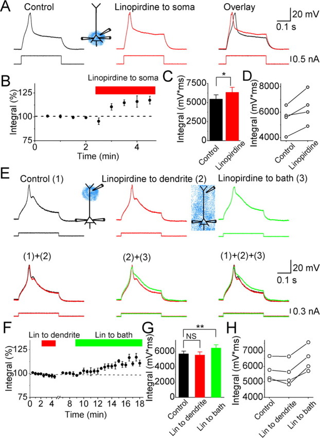
Effects of linopirdine on calcium spikes in CA1 pyramidal neurons. A, Typical example showing the effects of perisomatically applied linopirdine (30 μm; red) on the Ca2+ spike. B, Normalized time course of the effect of perisomatically applied linopirdine on the integral of the depolarizing response, including the Ca2+ spike. C, Summary diagram showing the effect of perisomatically applied linopirdine on the response integral (n = 4). D, The depolarizing response integral values from four cells before and after perisomatically applied linopirdine. E, Ca2+ spikes under control conditions [(1), black trace], after dendritic application of 30 μm linopirdine (212 μm from the soma) [(2), red], and then after bath applying 30 μm linopirdine [(3), green]. F, Normalized time course of the effect of dendritic focal application of linopirdine (Lin) and subsequent bath-applied linopirdine on the response integral. G, Summary diagram showing the response integral, under control conditions, after focally applying linopirdine to the dendrite, and then after bath applying linopirdine for all cells tested (n = 4). **p < 0.01; NS, not significant. H, Depolarizing response integral values from four cells under control conditions, after dendritic application of linopirdine, and subsequently after bath applying linopirdine. In all of the experiments illustrated in this figure, the hippocampal slices were incubated with 20 μm retigabine (to boost the M-current), 10 μm DNQX, 1 μm TTX, and 3 mm 4-AP, which were all routinely added to all the extracellular media. Ca2+ spikes were evoked by injecting a 300-ms-long depolarizing current pulse into the soma from a membrane potential of −68 mV.