
Figure 1.
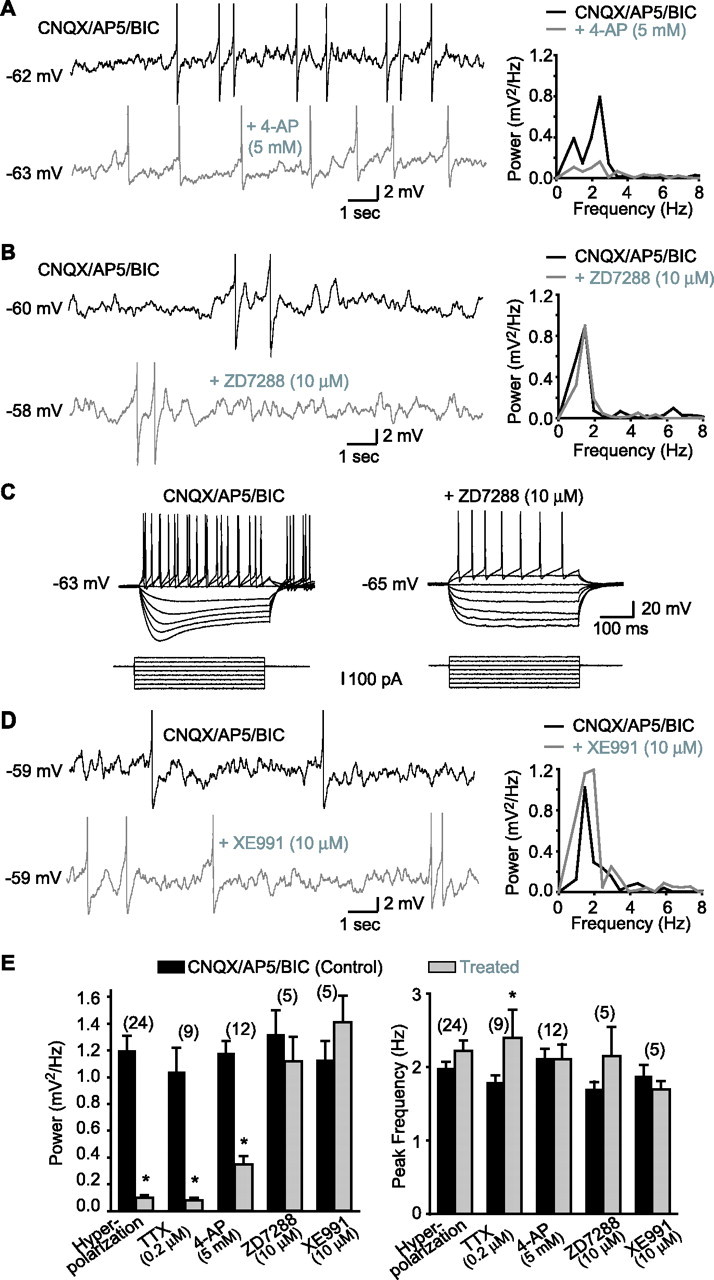
MPOs in LM/RAD interneurons in acute slices are dependent on 4-AP-sensitive K+ currents but not on Ih or IM. A, MPOs from a representative interneuron (left) recorded at membrane potential near spike threshold in the presence of non-NMDA, NMDA, and GABAA receptor antagonists [CNQX, 20 μm; AP5, 50 μm; bicuculline (BIC), 25 μm; top trace]. In the same cell, MPOs are significantly reduced by application of 4-AP (5 mm; bottom trace). In this and other figures with traces showing MPOs, action potentials are truncated. Power spectra (right) of records from the same cell show the reduction of the power of MPOs by 4-AP. B, Similar data from another cell showing that MPOs recorded near spike threshold are not diminished by ZD7288, a blocker of the h current (Ih). C, Positive control experiments from the same cell as in B showing that the sag in the membrane response elicited by hyperpolarizing current injections and produced by Ih (control in CNQX/AP5/BIC; left) was blocked by ZD7288 (10 μm; right). D, Representative traces from another interneuron showing that MPOs and corresponding power spectra are not reduced by XE991 (10 μm), a selective blocker of muscarine-sensitive K+ current (IM). E, Summary bar graphs showing that the power of MPOs was significantly reduced by membrane hyperpolarization (Vm threshold −10 mV), TTX (0.2 μm), and 4-AP (5 mm) but not by ZD7288 (10 μm) or XE991 (10 μm), whereas the peak frequency of MPOs was generally unchanged. *p < 0.05.