
Figure 1.
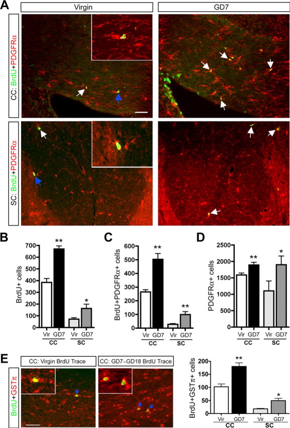
Pregnancy promotes increased OPC proliferation and the generation of new oligodendrocytes in the maternal CC and SC. A–D, Fluorescence micrographs (A) and quantification demonstrate increases in the number of BrdU+ cells (B), BrdU+PDGFRα+ cells (C; unpaired t test; CC, n =3; SC, n =5), and PDGFRα+ cells (D; unpaired t test; CC, n =7; SC, n =5) in the CC and SC of GD7 pregnant females relative to virgins (arrows indicate BrdU+PDGFRα+ cells; colabeled cells shown in insets are indicated with blue arrows). The image in the CC is taken in the body of the CC above the right ventricle, and the image of the SC is specifically of the dorsal funiculus. E, Fluorescence micrographs and quantification demonstrate increases in the number of newly generated oligodendrocytes (BrdU+GSTπ+ cells) in the CC (arrows indicate BrdU+GSTπ+ cell examples; colabeled cells shown in inset are indicated with blue arrows) and SC of GD7–GD18 BrdU trace animals relative to virgin trace animals 11 d after BrdU treatment (unpaired t test; CC, n =3; SC, n =5). Values are means ± SEM; *p < 0.05, **p < 0.01. Scale bars, 50 μm.