
Figure 2.
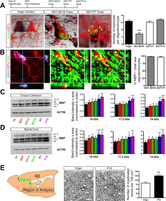
Newly generated oligodendrocyte maturation occurs during the postpartum period and is associated with increases in MBP levels and myelinated axons. A, Confocal z-stack images (3-D rendered images shown) and quantification of GSTπ+ processes (indicated by yellow arrows) extending from the soma of mature (GSTπ+; 60-d-old virgin animals) and newly generated oligodendrocytes (BrdU+GSTπ+) located in the CC of GD7–GD18, GD7–P7, and GD7–P14 BrdU trace pregnant animals (one-way ANOVA with Tukey's HSD post hoc test; n =3, N ≥ 25 cells per animal). Quantification suggests that new oligodendrocytes born on GD7 ultimately attain a normal complement of three to four processes by P7. B, Quantification using confocal imaging was used to determine the percentage of newly generated oligodendrocytes (BrdU+GSTπ+ cells) in the CC of GD7–GD18 and GD7–P7 BrdU trace animals (BrdU+GSTπ+ cell) that express MBP (n =3, N ≥ 25 cells per animal). The majority of new oligodendrocytes in the maternal CNS were observed to express MBP (arrows indicate yellow regions of coexpression in the z-axis under confocal microscopy; immunoreactivity was primarily observed within the processes rather than the cell soma). C, D, Western blot analysis of MBP expression in the CC (C) and SC (D) over the course of pregnancy and the postpartum period demonstrated significantly increased levels of the 18, 17.2, and 14 kDa MBP isoforms at P7 and P14 relative to virgin controls (one-way ANOVA with Dunnett's post hoc test; n =4 or more animals per group). E, Quantification of the number of myelinated axons in the genu of the CC in age-matched 11-week-old virgins (n =4) and P14 mothers using EM (unpaired t test; n =4) revealed a significant increase in the number of myelinated axons in the postpartum females. Values are means ± SEM; *p < 0.05, **p < 0.01, ***p < 0.001. Scale bars: A, 10 μm; E, 2 μm.