
Figure 3.
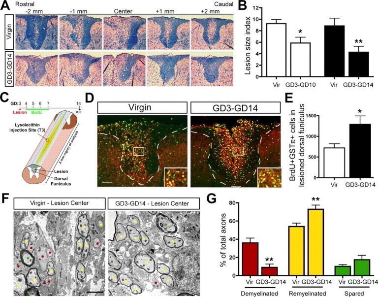
Pregnancy enhances the ability of the maternal CNS to regenerate demyelinating lesions. A, Luxol fast blue staining for myelin reveals the demyelinated lesion size in the SC dorsal funiculus over a 4 mm region of analysis in virgin and GD3–GD14 pregnant animals. B, Quantification of the proportion of the dorsal funiculus that remained demyelinated at 7 d (GD3–GD10, n =4; matched virgin control, n =4) and 11 d (GD3–GD14, n =8; matched virgin control, n =8) after lesion (unpaired t test) revealed a significant reduction in the lesion size of pregnant animals relative to matched virgin controls (Vir). C, Schematic overview of BrdU tracing experiment in lesioned virgin and GD3–GD14 pregnant animals and diagram showing lesion in center of 2 mm region of analysis. D, E, Fluorescent micrographs and quantification of BrdU+GSTπ+ cells in the lesioned dorsal funiculus of virgin (n =7) and GD3–GD14 pregnant animals (unpaired t test; n =7) demonstrating a significant increase in the number of newly generated oligodendrocytes in pregnant females relative to virgins. F, EM micrographs of demyelinated (red dots), remyelinated (yellow dots), and spared (green dots) axons within dorsal funiculus lesions of virgin and GD3–GD14 pregnant females. G, Quantification revealing the percentage of total axons that were demyelinated, remyelinated, or spared in virgin versus GD3–GD14 pregnant (unpaired t test; n =4) demonstrated that the proportion of demyelinated axons was reduced, whereas the proportion of remyelinated axons was increased in the pregnant females relative to the virgins. Values are means ± SEM; *p < 0.05, **p < 0.01. Scale bars: D, 100 μm; F, 2 μm.