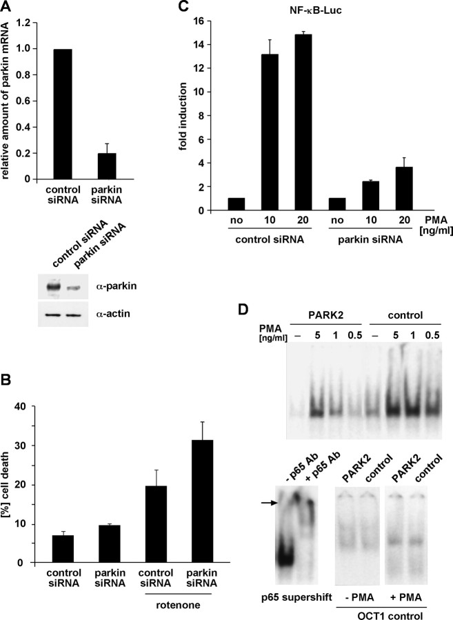
Figure 10.
Loss of endogenous parkin increases cell death and compromises NF-κB activation in response to stress. A, B, HEK293T cells were transfected with parkin-specific or control siRNA duplexes. A, At 48 h later, parkin mRNA levels were analyzed by RT-PCR (described in Fig. 1C), and endogenous parkin protein was analyzed by Western blotting (bottom) as described in Figure 1F. B, Parallel cultures were exposed to rotenone (0.05 μm, 3 h), and, after additional 24 h, cell viability was determined by trypan blue exclusion. C, HEK293T cells cotransfected with parkin-specific siRNA and the NF-κB reporter plasmid were incubated with PMA (10 and 20 ng/ml, 3 h). At 5 h later, cells were harvested and analyzed for luciferase activity. D, Fibroblasts from a PARK2 patient or control fibroblasts were exposed to PMA for 30 min. Nuclear extracts were prepared, and NF-κB binding activity was determined by electrophoretic mobility shift assays as described in Figure 2E. Bottom left, Supershift induced by a pAb against p65 in nuclear extracts from PMA-treated control fibroblasts. Bottom right, As a control, OCT1 binding activity was determined in nuclear extracts from untreated and PMA-treated PARK2 and control fibroblasts.

