
Figure 6.
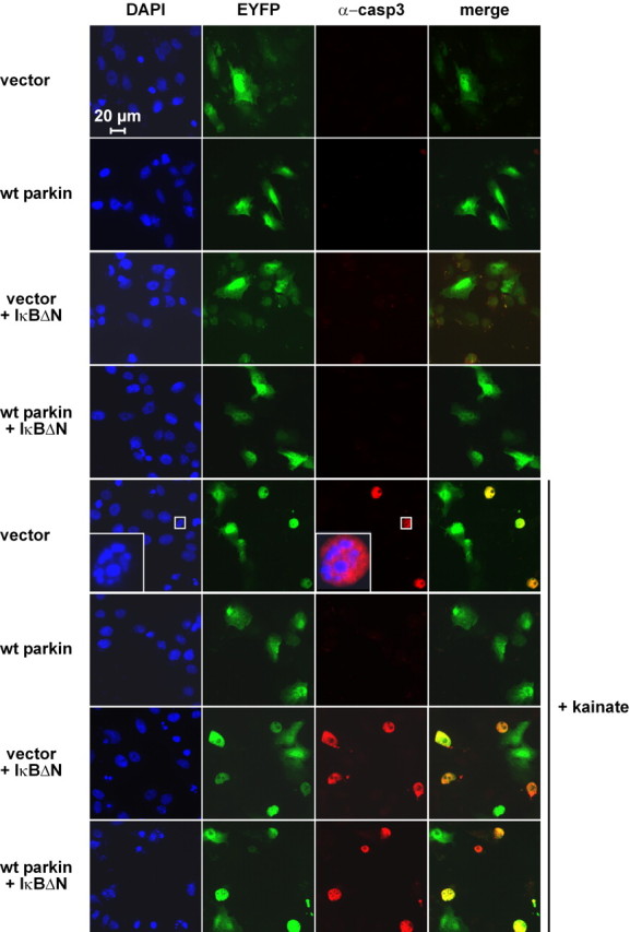
In the presence of the NF-κB super-repressor IκBΔN, parkin shows no neuroprotective activity. Immunofluorescence analysis of the experiment described in Figure 5A. Activation of caspase-3 was detected by using the anti-ACTIVE caspase-3 pAb (red), and nuclei were stained with DAPI (blue). Scale bar, 20 μm