
Figure 5.
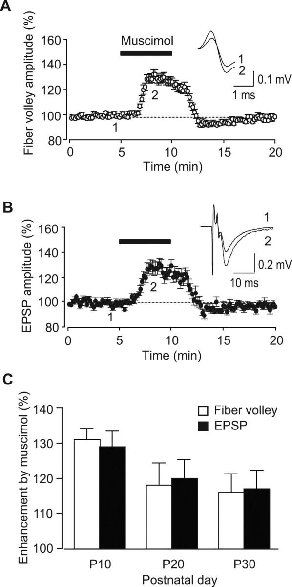
Muscimol-induced enhancement of fiber volleys and EPSPs at immature mossy fiber synapses. A, B, The time course of the amplitudes of fiber volleys (A) and EPSPs (B) before, during, and after the application of 0.1 μm muscimol at P10. In the inset, representative traces recorded at the times indicated by the numbers in the graph are shown. C, Developmental changes of the enhancement of fiber volley amplitudes (open columns) and EPSPs (closed columns).