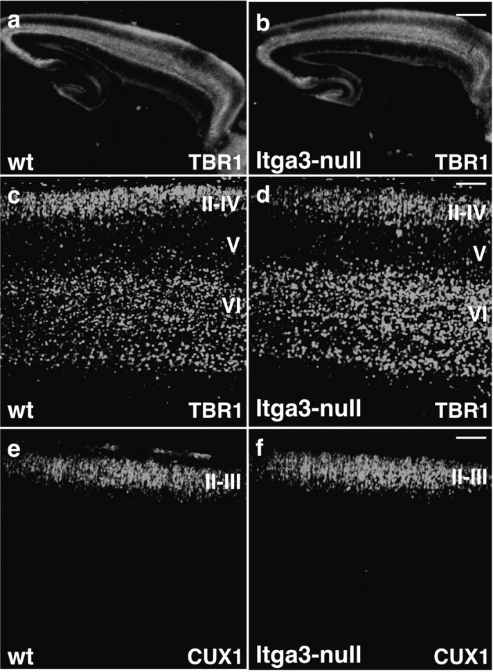
Figure 9.

Cortical layers form normally in Itga3-null mice. Sagittal sections through the cerebral cortex of wild-type and Itga3-null mice at P0 were stained with antibodies to TBR1, to reveal neurons in layers II–IV and VI (a–d); and CUX1, to reveal neurons in layers II–IV (e, f). Note that there was no difference in the distribution of different neuronal subtypes in wild-type and Itga3-null mice. Scale bars: a, b, 425 μm; c, d, 80 μm; e, f, 100 μm.
