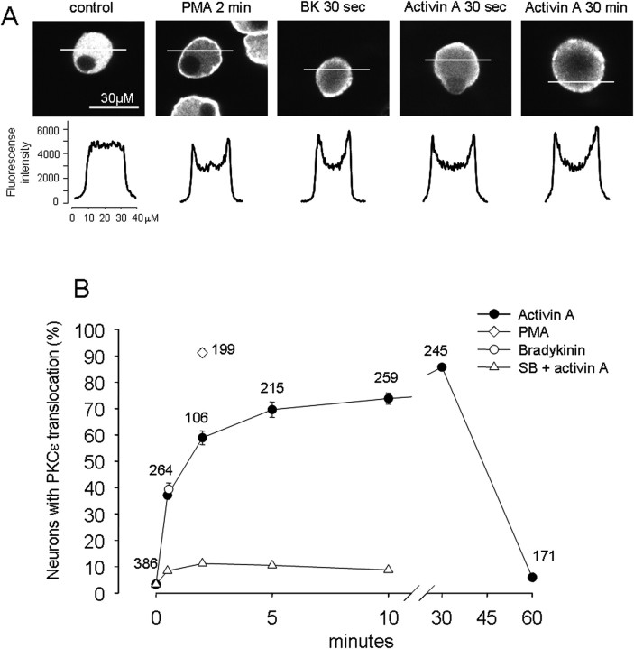
Figure 6.
Activin A causes translocation of PKCε to the cell membrane of rat DRG neurons. A, Distribution of PKCε immunoreactivity of rat DRG neurons after different treatment. Top, Confocal slice images of PKCε immunoreactivity under the indicated conditions. Bottom, Corresponding fluorescence intensity profile along a 40 μm line crossing each neuron. A uniform distribution of PKCε immunoreactivity was seen in the cytoplasm under control conditions, whereas a prominent relocation to the plasma membrane was seen for the conditions of PMA (300 nm, 2 min), BK (1 μm, 30 s), and activin (10 ng/ml, 30 s and 30 min). B, Time course of PKCε membrane translocation quantified as the percentage of neurons showing positive translocation. A positive neuron was taken as any neuron where the peak (membrane) fluorescence was ≥120% of the mean of 8 pixels in the center of the scan. At control time (no activin exposure), ∼3% of neurons showed PKCε membrane translocation, whereas at 30 s of activin exposure, ∼37% of neurons demonstrated PKCε membrane translocation, which peaked at ∼85% after 30 min. SB 431542 dramatically decreased PKCε membrane translocation. The potent PKC activator PMA (300 nm, 2 min) caused ∼91% of neurons to demonstrate PKCε membrane translocation, whereas BK (1 μm, 30 s) treatment triggered PKCε membrane translocation in ∼39% of neurons. Numbers of neurons measured are indicated for each data point.

