
Figure 7.
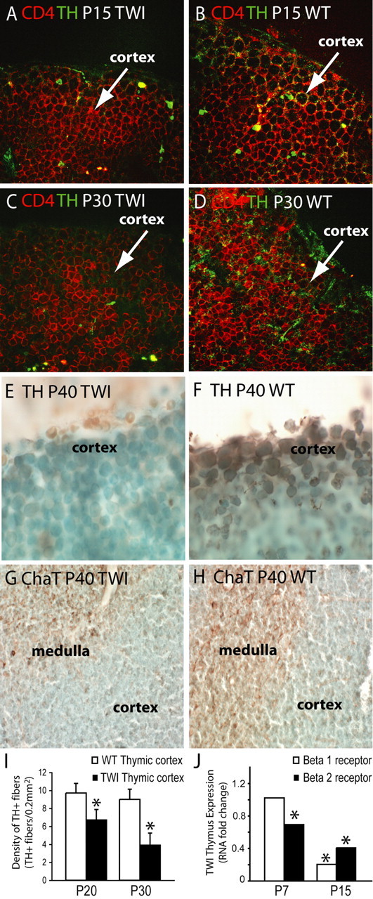
Progressive axonal loss from autonomic fibers in the atrophied GLD thymus. A–D, Confocal immunohistochemistry of TH (green) showed reduced innervation of norepinephrine fibers in the Twitcher (TWI) cortex at P15 (A) and P30 (C), when compared with age-matched controls (B, D). A CD4 counterstaining (red) was used. E–H, Light microscopy immunohistochemistry for TH (E, F) and ChaT (G, H) in P40 thymuses showed reduced expression of TH (E) and ChaT (G) in the aged TWI thymus. I, The density of TH+ fibers was quantified and expressed as the number of TH+ fibers per 0.2 mm2 of thymic cortex at P20 and P30. Data averages two independent experiments. p < 0.05 versus wild type. J, Fold changes in expression of norepinephrine receptors (β1 and β2) was quantified by real-time PCR in RNA isolated from thymuses at P7 and P15. Data are normalized to the expression of the GAPDH housekeeping gene and compared with values obtained in age-matched wild-type tissues (the expression of which equals a value of 1). Data average two independent experiments. p < 0.05 versus wild type.