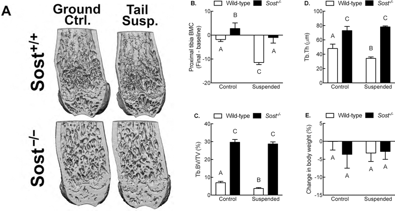
Figure 5. Sost is required for bone wasting under disuse conditions.
(A) Representative μCT reconstructions of the distal femur from ground control and tail-suspended wild-type and Sost−/− mice. (B) Percent change (pre-suspension scan vs. post-suspension scan) in proximal tibia bone mineral content from ground control and tail-suspended wild-type and Sost−/− mice as measured by serial pQCT scans. Bone volume fraction (C) and trabecular thickness (D) in the distal femur metaphysis as measured by μCT. (E) Percent change (pre-suspension vs. post-suspension) in body mass in ground control and tail-suspended wild-type and Sost−/− mice. *p<0.05 for tail suspended mice vs genotype-matched control mice. Different letters denote statistical differences among groups. n = 8–10/group.

