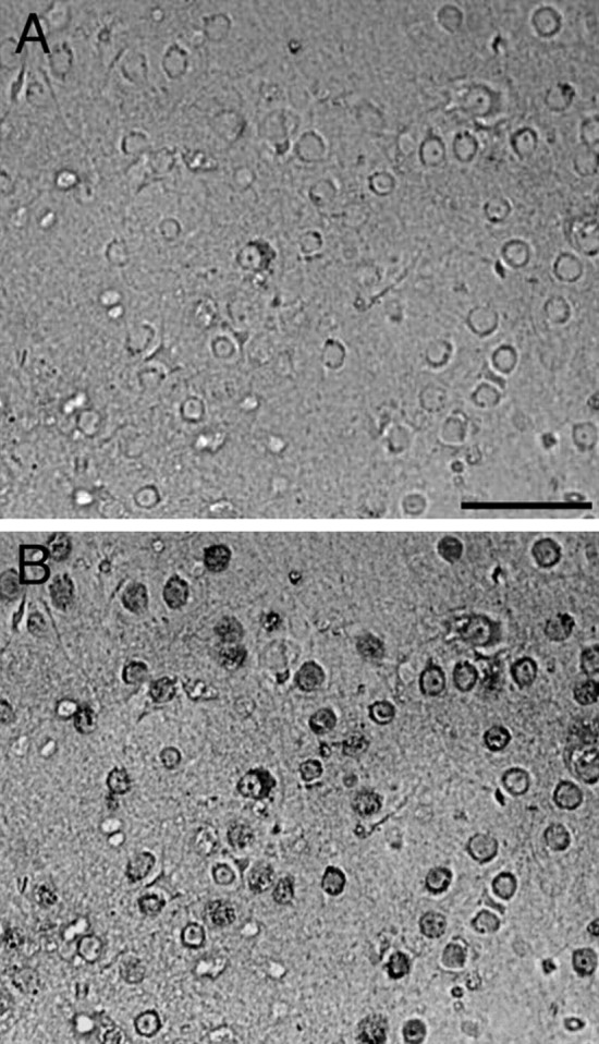
Figure 3.

Neuronal preparation from neonatal rat cortical cells. A, Phase contrast photomicrograph of the neuronal preparation. B, Immunostaining of the same field with mouse anti-NeuN (dark gray) examined with fluorescent microscope. Scale bar, 30 μm
