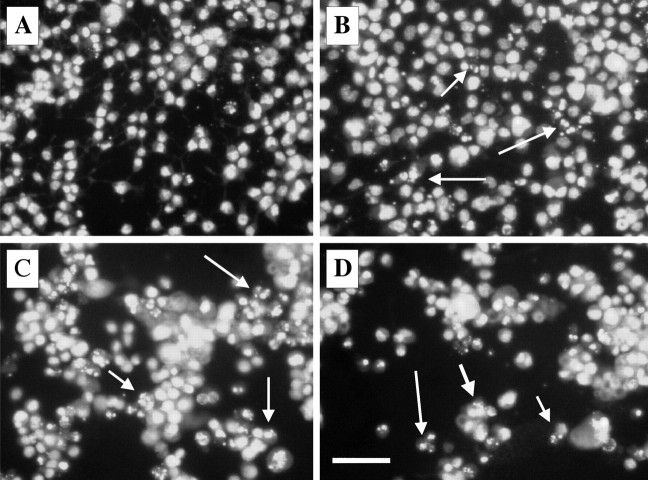
Figure 8.
Time course of the PrP 82–146 induced nuclear fragmentation of N2a cells. N2a cells (A, control condition) were exposed to PrP 82–146wt for 24 h (B), 48 h (C), and 96 h (D). Cells were analyzed by fluorescence microscopy after staining with the DNA-binding fluorochrome (Hoechst 33258). The percentage of fragmented nuclei progressively increased with time. The arrows indicate nuclei with typical apoptotic features. Scale bar, 20 μm.

