
Figure 1.
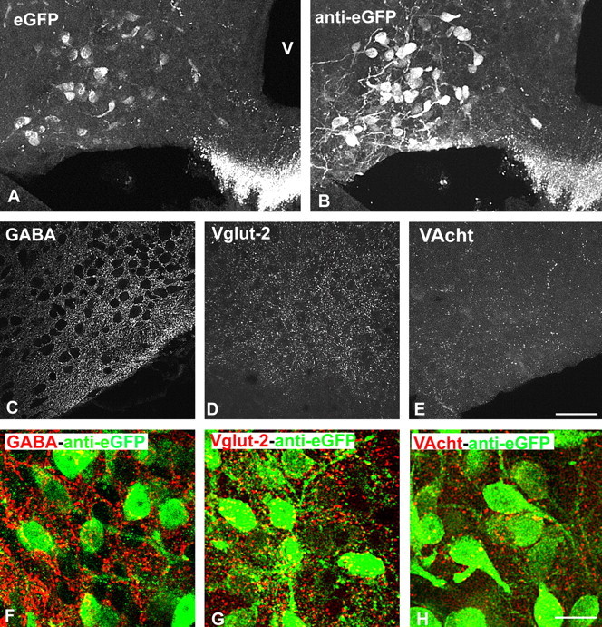
Visualization of GHRH neurons and their putative afferent fibers in the arcuate nucleus of GHRH-GFP mice. A, B, GHRH-GFP neurons before (A) and after (B) eGFP immunostaining. Pictures are taken from the same brain section. C–E, Immunocytochemical visualization of GABAergic (anti-GABA, C), glutamatergic (anti-VGLUT2, D), and cholinergic (anti-VACht, E) structures within the arcuate nucleus. Axon terminal-like structures are dispersed within the arcuate nucleus. F–H, Double immunocytochemical labeling of eGFP and of other neurotransmitters systems. eGFP-labeled cell bodies and dendritic-like processes are frequently surrounded by GABA- and VGLUT2-labeled axons, but only rarely by VACht-labeled axons. Pictures are from male mice. V, Third ventricle. Scale bars: (in E) A–E, 50 μm; (in H) F–H,15 μm.