Figure 4.
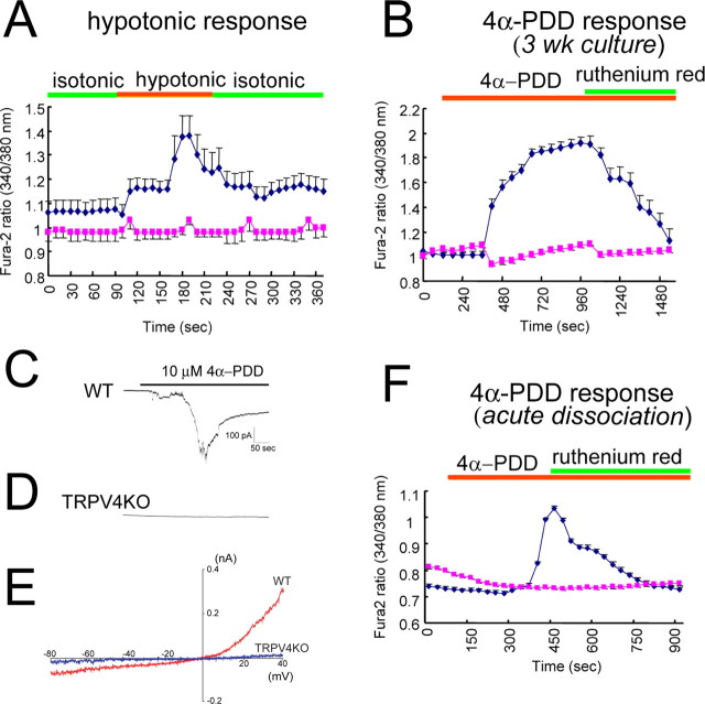
Functional TRPV4 is expressed in hippocampal neurons. A, Averaged traces of [Ca2+]i changes (with SEM) on hypotonic stimulus (250 mOsm/kg from isotonic condition; 322 mOsm/kg) in WT (blue) and TRPV4KO (red) neurons (21 DIV; 4 independent experiments; n = 156–193) at 25°C. B, Averaged traces of [Ca2+]i changes with 4α-PDD (10 μm) application in WT (blue) and TRPV4KO (red) neurons (21 DIV; 4 independent experiments; n = 161–199) at 25°C. Ruthenium red (10 μm) almost completely inhibited 4α-PDD-evoked [Ca2+]i increase. C, D, Representative current traces on 4α-PDD application in WT (C) or TRPV4KO (D) neurons (21 DIV). Holding potential was –60 mV. E, Representative current–voltage relationship of 4α-PDD-evoked currents in WT (red) and TRPV4KO (blue) neurons (21 DIV). F, Averaged traces of [Ca2+]i changes on 4α-PDD (10 μm) application in acutely dissociated neurons from WT (blue) and TRPV4KO (red) mice (4 independent experiments; n = 814) at 25°C. Ruthenium red (10 μm) almost completely inhibited 4α-PDD-evoked [Ca2+]i increase. Green and orange bars in A, B, and F indicate application time of the indicated stimuli or compounds.

