
Figure 10.
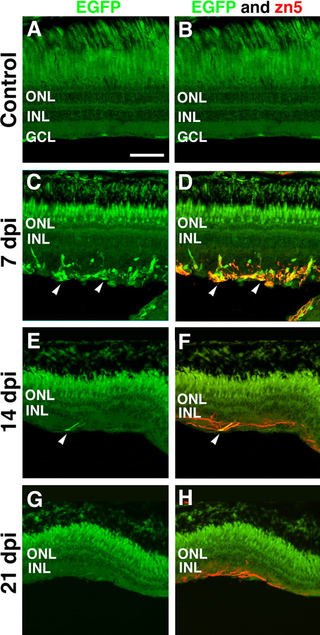
Frozen retinal sections of the Tg(atoh7:EGFP)nt transgenic zebrafish were used to assess the temporal expression of atonal homolog 7 and zn5 during regeneration of the ouabain-damaged retina. The left column shows EGFP immunolocalization in the Tg(atoh7:EGFP)nt transgenic retina, and the right column shows coimmunolocalization of EGFP and zn5 (in red). A, B, Uninjected control retinal sections did not reveal either EGFP (green) or zn5 expression (red). C, D, At 7 dpi, EGFP expression was localized to several scattered cells in the regenerating inner retina, and several of these cells colocalized with zn5 immunoreactivity (arrowheads). E, F, At 14 dpi, EGFP and zn5 coexpression was restricted to a limited number of ganglion cell axons found in the inner retina (arrowheads). G, H, At 21 dpi, all of the retinas examined lacked EGFP expression. However, zn5 immunoreactivity persisted on axonal projections in the regenerating inner retina. Scale bar, 75 μm.