
Figure 4.
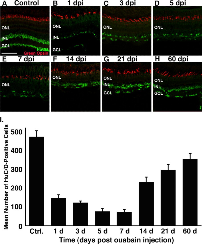
Immunohistochemical analysis of HuC/D and green opsin expression in the ouabain-treated retina. Frozen retinal sections were double immunolabeled with anti-HuC/D monoclonal antibody (green), to detect differentiated amacrine and ganglion cells, and rabbit anti-green opsin polyclonal antiserum (red), a marker for one member of the double cone cell pair. A, Control section from a wild-type retina injected with 0.65% saline possessing HuC/D-positive cells in the GCL and the distal portion of INL and well defined green cone cell outer segments. B–E, During the first 7 dpi, the number of HuC/D-positive cells decreases significantly, and the distinction between the INL and GCL is lost. Green cone cells, however, maintain morphological integrity and relatively normal levels of green cone opsin. F–H, After 7 dpi, there is a significant increase in the number of HuC/D-positive cells and reestablishment of retinal lamination. I, Graph of the number of HuC/D-positive cells in the dorsal retina (y-axis) versus the days after ouabain injection (x-axis). Ctrl., Control. Error bars represent SD (n = 4 retinas per time point). Scale bar, 75 μm.