
Figure 5.
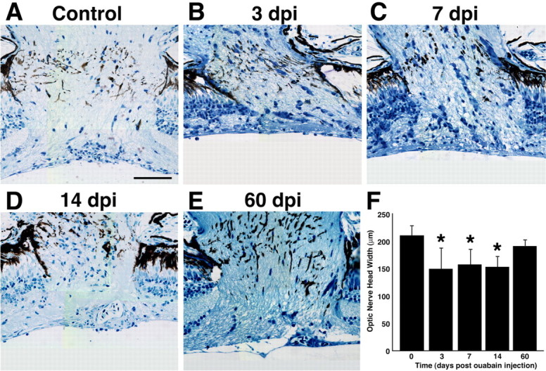
Histological time course of the diameter of the optic nerve head in ouabain-treated eyes. A–E, Images were collected from central optic nerve sections from a wild-type uninjected control retina (A) or retinas injected with 2 μm ouabain (B–E). B, At 3 dpi, the extensively damaged retina (Fig. 2C) retains axonal fibers in the optic nerve head. B–D, Although the optic nerve head was significantly reduced in diameter from 3 to 14 dpi relative to the control, axonal fibers can be clearly identified in all histological sections. E, At 60 dpi, the ouabain-damaged retina possesses an optic nerve head diameter that is statistically similar to the uninjected control retina. F, Graph of the optic nerve head width (y-axis) versus the days after ouabain injection (x-axis). Three measurements were made across the width of the optic nerve head in each section, with two sections analyzed for each eye. Error bars represent SD (n = 6 retinas per time point). Asterisks denote values that are statistically different from the control (p ≤ 0.05). Scale bar, 50 μm.