
Figure 5.
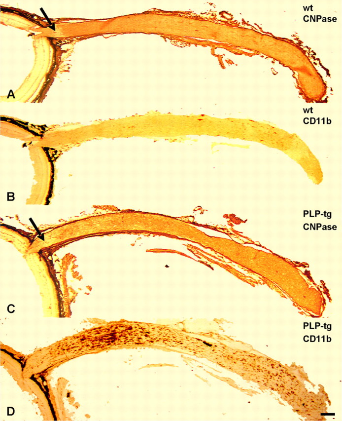
Macrophage-like cells in the optic nerves of wild-type and PLP mutants. A–D, Longitudinal, consecutive frozen sections of retinas (left) and optic nerves from wt mice (A, B) and PLP mutants (PLP-tg; C, D) immunolabeled with antibodies to CNPase (A, C) and with antibodies to the macrophage-specific marker CD11b (B, D). Note that particularly in the rostral part of the mutant optic nerve, there is a substantially increased number of CD11b+ cells compared with the optic nerves of the wild-type mice. In this region, myelin damage is particularly prominent, as reflected by a more patchy CNPase labeling. Arrows in A and C mark the unmyelinated part of the optic nerve, which is clearly confined by myelin labeling in the wild-type mice but diffusely in the mutants. Labeling of meninges and connective tissue of the eye cup in A and C is attributable to unspecific binding of the corresponding secondary antibody. The black appearance associated with the eye cup in all micrographs corresponds to the pigment epithelium. Scale bar, 200 μm.