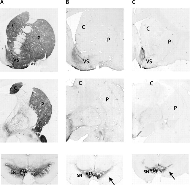
Figure 1.
Photomicrographs of TH staining demonstrating the loss of dopaminergic substantia nigra pars compacta neurons in the MPTP-treated monkeys compared with a control animal. A was taken from a control normal macaque monkey; B and C are from the MPTP-treated monkeys R (macaque) and Q (vervet), respectively. The photomicrographs illustrate the levels of rostral striatum (row 1), central striatum (row 2), and midbrain (row 3). Note the lack of TH-positive staining throughout the striatum with the exception of the ventral striatum, particularly the shell region. TH-positive cells are selectively lost in the ventral tier (see arrows) but selectively spared in the ventral tegmental area. C, Caudate; P, putamen; VS, ventral striatum; SN, substantia nigra; VTA, ventral tegmental area.

