Figure 3.
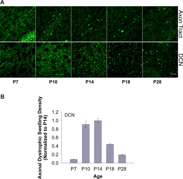
Formation of GFP–LC3-labeled axonal dystrophic swellings as an early response in the degenerating Purkinje cells of Lurcher mice. A, Representative confocal images show the development of GFP–LC3-labeled axonal dystrophic swellings in axon tracts of cerebellar white matter (top row) and in the DCN (bottom row) of degenerating Purkinje cells of GFP–LC3/Lurcher mice at different postnatal days. Scale bar, 20 μm. B, Quantification of the images in A shows that the density of axonal dystrophic swellings in the DCN of the degenerating Purkinje cells of GFP–LC3/Lurcher mice (normalized to P14) was at peak level between P10 and P14. The numbers of images used for quantification at the ages of P7, P10, P14, P18, and P28 were 13, 14, 16, 7, and 5, respectively. Error bars indicate SEs.

