
Figure 7.
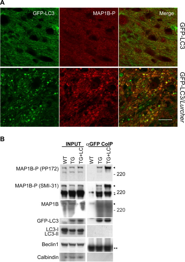
Increased amount of MAP1B-P bound to GFP–LC3 in axonal dystrophic swellings of GFP–LC3/Lurcher Purkinje cells. A, Representative confocal images of a cerebellar slice of GFP–LC3/Lurcher mouse (P10) immunostained with anti-MAP1B-P antibody (PP172) show partial colocalization of GFP–LC3 (in green) and MAP1B-P (in red) in the axonal dystrophic swellings of degenerating Purkinje cells. Scale bar, 20 μm. B, Western blot analysis of cerebellar tissue extracts (INPUT) from wild-type (WT), GFP–LC3 transgenic (TG), and GFP–LC3/Lurcher mice (TG+LC) and their anti-GFP immunoprecipitations (αGFP CoIP) shows increased binding of MAP1B-P, but not MAP1B, to GFP–LC3 in GFP–LC3/Lurcher cerebellum compared with GFP–LC3 transgenic cerebellum. Negative controls show the lack of endogenous LC3, beclin 1, or calbindin in the anti-GFP immunoprecipitations.