
Figure 2.
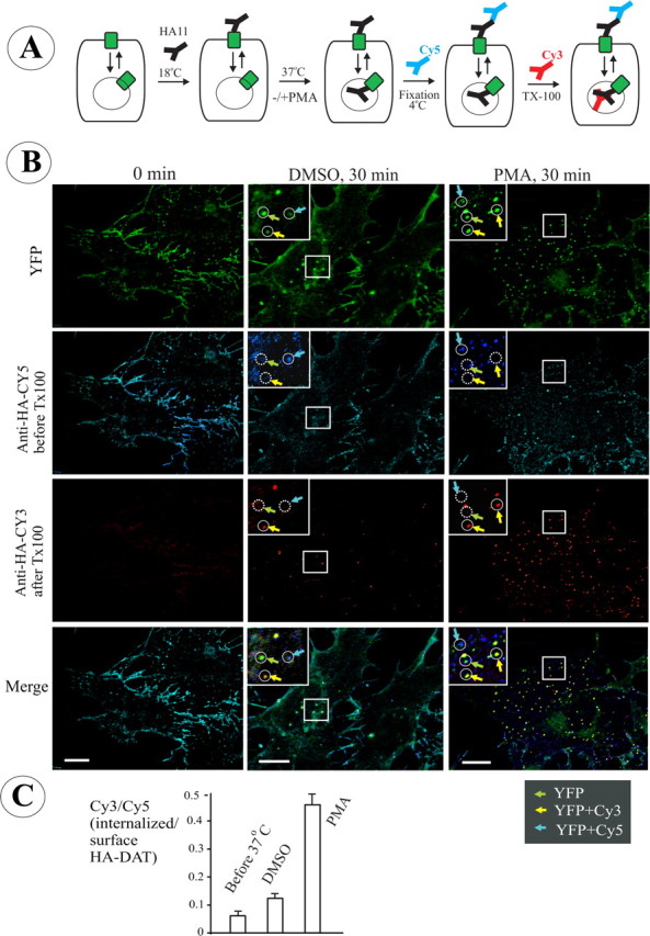
Single-cell endocytosis assay using HA11 antibody in PAE cells expressing YFP-HA-DAT. A, Schematics of the endocytosis assay using differential staining of surface and internalized HA11. The cells were incubated with HA11 at 18–22°C for 30 min, washed, further incubated at 37°C, and fixed. The nonpermeabilized cells were then stained with Cy5-tagged anti-mouse secondary IgG to occupy all surface HA11/YFP-HA-DAT complexes. After Triton X-100 (TX-100) permeabilization, the cells were incubated with the same secondary antibody but tagged with Cy3 to mark internalized HA11. B, Representative images of PAE/YFP-HA-DAT in cells treated with vehicle (DMSO) or PMA (1 μm) for 30 min at 37°C after prebinding of HA11. The staining with Cy5 (blue) and Cy3 (red) was performed as described in A. The sum projection images of four consecutive optical sections are presented. Examples in high-magnification insets (single optical sections) demonstrate clusters and compartments containing YFP alone (total YFP-HA-DAT; green arrows), YFP colocalized with Cy5 (plasma membrane HA11; cyan arrows), and YFP-colocalized with Cy3 (internalized HA11; yellow arrows). Identical positions in the insets are indicated by circles. Solid and dashed lines show, respectively, the presence and absence of an endosome or a plasma membrane cluster within the circle. Scale bars, 10 μm. C, Quantification of the ratio of internalized/surface YFP-HA-DAT (mean ± SD; n = 8–9 cells) from the images presented in B (for details, see Results).