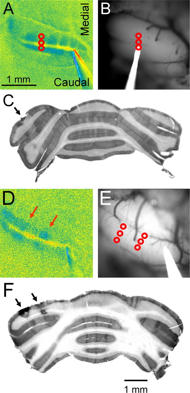
Figure 5.

Bands of decreased fluorescence are in register with the zebrin II-positive band. A, Response to surface stimulation and the site of three electrolytic lesions (red circles) placed between two inhibitory bands in the center of the folium. B, Background image of the folium showing the lesion sites and the lesion electrode in place. C, Anti-zebrin II immunostaining of a section through the folium shows the location of the lesions between two zebrin II bands that correspond to P5b+ and P6+ (Eisenman and Hawkes, 1993; Sillitoe and Hawkes, 2002). D–F, Similar series of images in which two sets of lesions were placed within the two inhibitory bands (D). The recovered lesions are located on the P5b+ and P6+ zebrin II bands (F).
