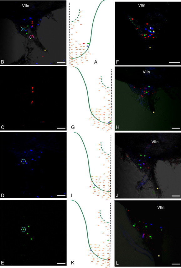
Figure 3.

Injecting three adjacent buds with different color markers results in three separate groups of labeled neurons in the geniculate ganglion. A, Three adjacent buds, RIV-1, RV-1, and RV-2, were injected with DiD (blue), DiO (green), and DiI (red), respectively. B, Labeled ganglion cells from three buds in A, viewed with all three fluorescence settings, are predominantly single labeled and scattered in the ganglion. A double-labeled cell from RIV-1 and RV-1 injections is marked by the cyan circle (same cell circled in D and E); cells encircled in pink are two separate cells from RIV-1 and RV-2 buds as determined by confocal layer analysis. C–E, Cells innervating each bud imaged separately with the appropriate laser settings. G, I, Three adjacent buds injected with different color markers in two different regions show separate labeling of all ganglion cells (F, H, and J, respectively). K, Three widely separated buds (CI-6, CV-3, and CI-16) injected with the different markers. L, Separate labeling of ganglion cells. The asterisk marks the GSP nerve. VIIn marks the seventh cranial nerve. Scale bars, 100 μm.
