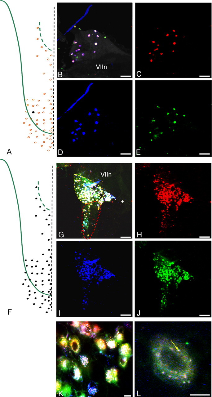
Figure 5.

A–E, Injecting a single bud (RIII-2) with an equimolar mixture of the three dyes used in the present study (DiI, DiD, and DiO), a control for the equal effectiveness of dye uptake, labeled all of the same ganglion cells with every marker. A, Injection site. B, Merged image of all three colors showing all triple-labeled cells (white containing). The pink cast of some cells reflects the variable intensity of labeling because of physical chemistry of dye miscibility at the injection site. The blue line and green patches are artifacts. C–E, Separate images of each marker viewed with the appropriate fluorescence settings. F–L, All chorda tympani nerve fibers innervating taste buds (F) were labeled with an equimolar mixture of the three dyes. G, Most ganglion cells in the merged image of all three colors were triple labeled. H–J, Separate images of each marker viewed with the appropriate settings. K, L, Higher-magnification view showing that each marker could be discerned inside the cells as different colored vesicles, some of which show precise overlap of all three colors (white puncta at arrow in L). Scale bars: B–E, G–J, 100 μm; K, L, 10 μm. The asterisk marks the GSP nerve, whereas the + symbol marks the central (toward brain) corner of the ganglia. VIIn marks the facial nerve.
