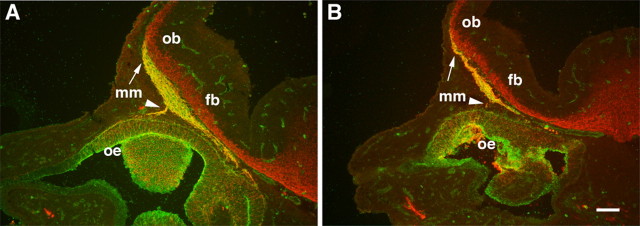
Figure 7.
The MM is abnormal in CXCR4−/− mice. Double-label immunofluorescence analysis of sagittal sections through the nose and forebrain of E12 mice with the use of anti-βIII-tubulin (red) antibodies and FITC-conjugated LEA lectin (green) labels the MM (yellow). A, In CXCR4+/− mice double-labeled (yellow) MM cells and axons extend from the OE (arrowhead) to the rostral tip of the OB (arrow), which has begun to protrude from the forebrain (fb). B, In a matched section through the nose and forebrain of a CXCR4−/− mouse, the MM is significantly smaller, containing many fewer double-labeled (yellow) cells and axons. In contrast, βIII-tubulin expression in the forebrain appears normal in CXCR4−/− mice. Scale bar, 100 μm.

