
Figure 1.
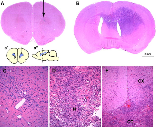
PDGF overexpression induces the formation of malignant gliomas. Hematoxylin and eosin stains were performed on coronal sections of adult rat brains 14 dpi with pQ-GFP (A, C) or PDGF-IRES-GFP (B, D, E). A, No tumors formed in brains injected with the control retrovirus, but a small area of reactive gliosis is seen around the needle track (arrow). C, Higher-magnification micrograph showing the injection site. B, Large infiltrative tumors with the histological features of human glioblastoma formed by 14 dpi with PDGF-IRES-GFP retrovirus. This section, 3 mm caudal from injection site, shows the tumor extending across the corpus callosum into the contralateral hemisphere. D, Higher-magnification micrograph showing an area of pseudopalisading necrosis (N), a hallmark of glioma malignancy seen in human glioblastomas. E, Tumor cells crossing the corpus callosum (CC) and infiltrating the cortex (CX). Scale bar: (in B) A, B, 2 mm. Insets in A are coronal (a′) and sagittal (a″) schematic diagrams of the injection site (arrow) at the level of the forceps minor corpus callosum. The dotted line in a″ shows the level of the coronal section shown in B.