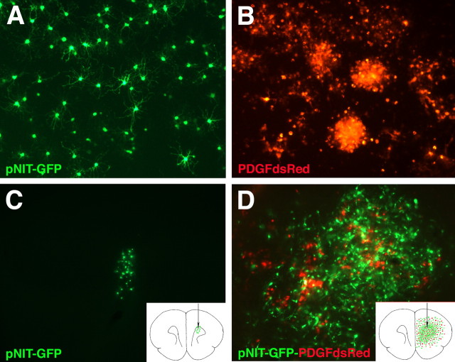
Figure 8.
Adult white matter progenitors infected with PDGF-IRES-DsRed in vitro form malignant gliomas through autocrine and paracrine signaling. Normal adult white matter progenitors were isolated and expanded for 5 d in vitro with B104-containing media. Proliferating progenitors were then infected with pNIT-GFP or PDGF-IRES-DsRed. A, B, Adult white matter progenitors grown in culture for 10 dpi in basal media. A, pNIT-GFP-infected cells stopped proliferating and acquired a highly branched morphology. B, PDGF-IRES-DsRed-infected cells retained a simple morphology and remained highly proliferative, forming large clusters of cells. C, D, At 2 d postinfection, the cells were implanted into adult brains. Brains were analyzed at 20 dpi. C, pNIT-GFP-infected cells injected alone remained close to the injection site and no tumors formed. D, When pNIT-GFP-infected cells were coinjected with PDGF-IRES-DsRed-infected cells, a tumor formed by 20 dpi, and this tumor is composed of a mixture of red and green cells. Equal numbers of pNIT-GFP cells were injected in C and D. Note the marked proliferation and dispersion of pNIT-GFP cells in the presence of PDGF-IRES-DsRed cells (D). Insets in C and D are schematic diagrams illustrating the distribution and number of cells (green and red dots) in coronal sections of adult brain at the level of the injection site.

