
Figure 2.
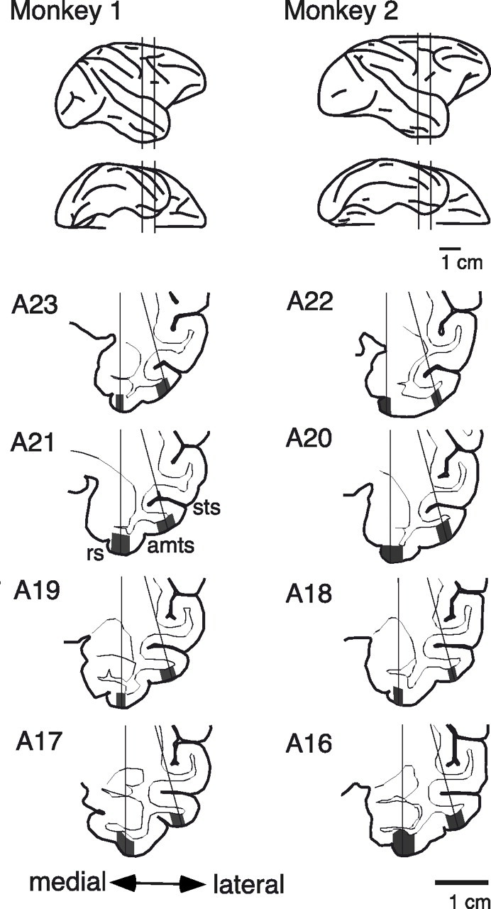
Recording positions shown in lateral and ventral views (top two lines) and frontal sections (bottom four lines) of the brain. The vertical lines in the lateral and ventral views indicate the posterior and anterior limits of recordings. The gray regions in the frontal sections indicate areas from which the recordings were made. The medial regions were within the perirhinal cortex, and the lateral regions were in area TEad. The recordings were conducted in the right hemisphere in both monkeys. sts, Superior temporal sulcus; amts, anterior middle temporal sulcus; rs, rhinal sulcus. The numbers to the left of the drawings indicate the distance (in millimeters) of the section from the ear bar position.