Figure 3.
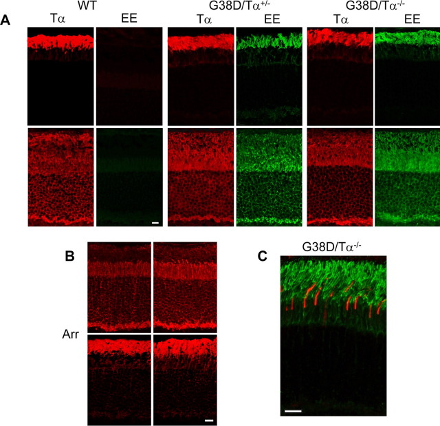
A, Localization and light-dependent translocation of Tα and G38D. Cryosections of the mouse retinas were obtained from dark-adapted (top row) and light-adapted (bottom row) mice (light adaptation: 50 min, ∼200 lux). The sections were stained with rabbit anti-rodTα antibody T1A or a monoclonal anti-EE antibody (Covance) and visualized with goat anti-rabbit AlexaFluor 568 and goat anti-mouse AlexaFluor 488 secondary antibodies using a Zeiss LSM 510 confocal microscope. WT, Wild type. B, Light-dependent translocation of arrestin (Arr). Cryosections of the mouse retinas from dark- and light-adapted mice (light adaptation: 50 min, ∼200 lux) were stained with rabbit anti-arrestin antibody and visualized with goat anti-rabbit AlexaFluor 568 secondary antibodies using a Zeiss LSM 510 confocal microscope. C, G38D is not expressed in cone photoreceptor cells. Cryosection of mouse retina from a dark-adapted G38D/Tα−/− mouse was double stained with rabbit anti-coneTα antibody and a monoclonal anti-EE antibody (Covance) and visualized with goat anti-rabbit AlexaFluor 568 and goat anti-mouse AlexaFluor 488 secondary antibodies using a Zeiss LSM 510 confocal microscope. Scale bars: A, B, 20 μm; C, 10 μm.

