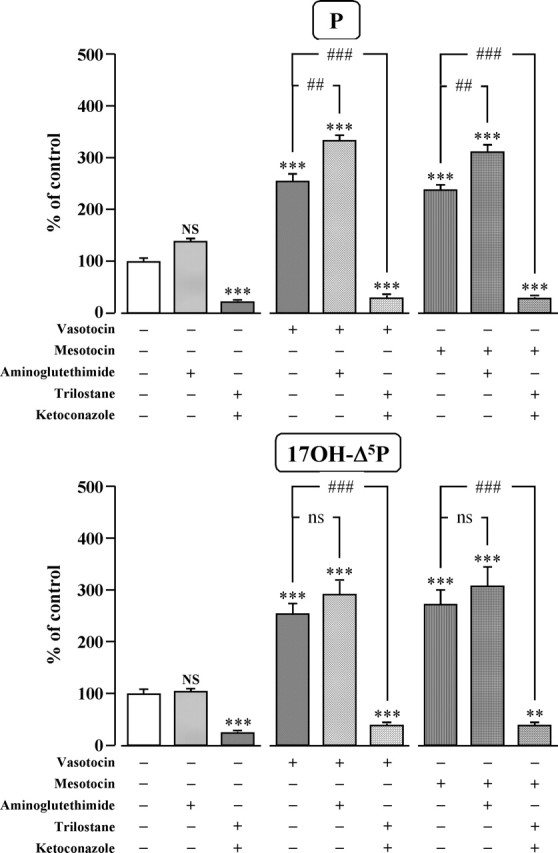
Figure 8.

Effects of VT (10−7 m) and MT (10−7 m) in the absence or presence of the cytochrome P450scc inhibitor aminoglutethimide (10−4 m), and the 3β-HSD inhibitor trilostane (10−4 m) and cytochrome P450C17 inhibitor ketoconazole (10−4 m) on the conversion of tritiated pregnenolone into P and 17OH-Δ5P by frog hypothalamic explants. The values were calculated from the areas under the peaks in chromatograms similar to those presented in Figure 5. Results are expressed as percentages of the amount of each steroid formed in the absence of drugs (control). Each value is the mean ± SEM of four independent experiments. ***p < 0.001 versus control; ##p < 0.01, ###p < 0.001 versus VT- or MT-stimulated levels; NS, not statistically different from control; ns, not statistically different from VT- or MT-stimulated levels (one-way ANOVA followed by a post hoc Bonferroni’s test).
