Figure 2.
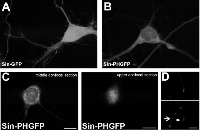
The PLCγ1PH domain is associated to intracellular and extracellular membranes. A, B, Single confocal planes of dissociated hippocampal cells transduced by Sin–GFP (A) or Sin–PHGFP (B). The PHGFP domain shows a characteristic patchy distribution of a membrane associated protein, whereas Sin–GFP is evenly distributed in the cytoplasm and the nucleus. C, The PLCγ1PH domain is associated to intramembrane and plasma membrane. Two individual confocal planes of one hippocampal neuron expressing the GFP-tagged PLCPH domain are shown. In the middle plane, a clear accumulation in intracellular membranes is visible, whereas a plane through the bottom of the cell demonstrates the accumulation of the protein in the plasma membrane. D, The PLCγ1PH domain is expressed and transported into tips of axons (arrowhead) and dendrites (arrow) already 6 h after Sindbis viral-mediated transduction. In the top panel, the staining with the axonal marker tau-1 is shown, and in the bottom panel, the patchy GFP fluorescence of the GFP-tagged PH domain is shown. Scale bars, 10 μm.

