
Figure 1.
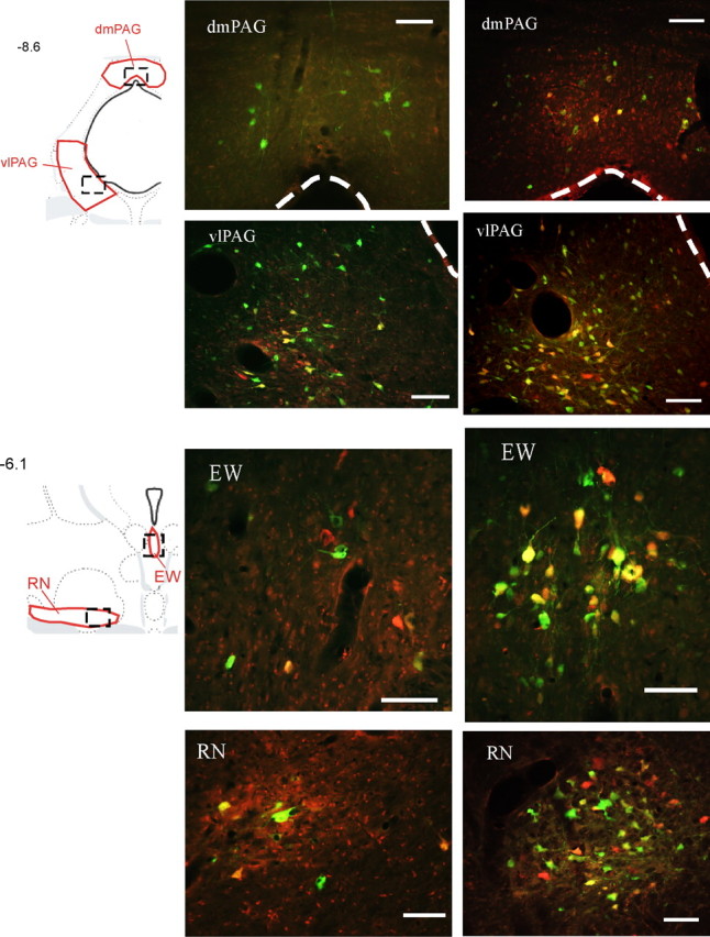
Examples of sympatho-motor neurons in the midbrain. Transsynaptically labeled neurons infected with PRV-152, which was injected into the gastrocnemius muscle, appear green, whereas those infected with PRV-BaBlu were pseudocolored with red; double-infected neurons appear yellow or orange. Drawings in the left column are from Swanson's rat atlas (Swanson, 2004) and indicate locations of anatomical regions shown to the right. Areas shown in red indicate boundaries that were used for cell quantifications, whereas boxes drawn with dashed lines indicate approximate locations of images on the right. Images in the middle column were taken from an animal that survived 132 and 108 h after injections with PRV-152 and PRV-BaBlu, respectively. Images in the right column are from an animal that survived 144 and 120 h after injections with PRV-152 and PRV-BaBlu, respectively. Numbers on the left indicate approximate distances from bregma in millimeters. dmPAG, Dorsomedial periaqueductal gray. Scale bars, 100 μm.