
Figure 7.
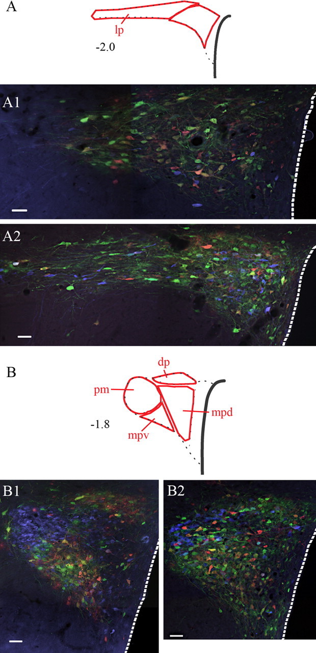
Relationship of sympatho-motor neurons to AVP- and OT-containing neurons in the PVN. Sections were processed for simultaneous visualization of viral reporter proteins (β-gal and eGFP) as well as AVP (A1, B1) or OT (A2, B2). β-Gal and eGFP were tagged with red and green fluorophores, whereas AVP and OT were tagged with a blue fluorophore. Note the extensive interdigitation of AVP-positive (A1, B1) and OT-positive (A2, B2) neurons with virally infected cells. Also note the differences in the distribution of AVP and OT within the PVN. Although AVP-positive neurons are found within all of the different subdivisions of the PVN, these cells are enriched in the posterior magnocellular (pm) subdivision (B1). In contrast, OT-containing cells show only light clustering within the pm subdivision but instead are scattered throughout the PVN (A2, B2). Abbreviations are as in Figure 3. Scale bars, 50 μm.