Figure 1.
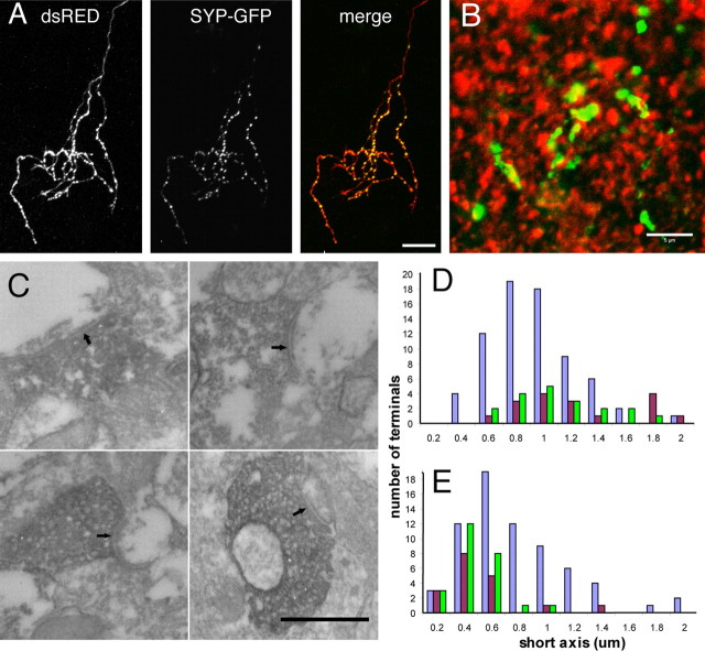
SYP-GFP labels presynaptic sites in vivo. A, Coexpression of SYP-GFP with dsRED-MST or SYP-CFP with EYFP in RGCs reveals discrete SYP puncta concentrated in the axon arbor in the optic tectum. These colocalize with the varicosities normally observed at regular intervals in the axon arbor. B, Immunohistochemistry for the postsynaptic marker PSD-95 (red) in the optic tectum of st48 tadpoles resulted in a punctate pattern that labeled sites directly juxtaposed to SYP-GFP (green) puncta in RGC axons. C, Electron microscopy examination of RGC terminals expressing the HRP-SYP-GFP fusion protein reveals dark DAB reaction product in synaptic vesicle membranes at morphologically identifiable synaptic sites (arrows). D, The distribution of presynaptic terminal sizes in RGC axons expressing the HRP-SYP-GFP (green) was indistinguishable from that in biotin dextran-labeled RGC axons (brown). Both were larger than the presynaptic terminals of unlabeled (nonretinal) synapses (purple) in the optic tectum. E, The contacted postsynaptic profiles were also identical for HRP-SYP-GFP-labeled RGC terminals and control biotin dextran-labeled RGC axons. Scale bars: A, 10 μm; B, 5 μm; C, 500 nm.

