Figure 1.
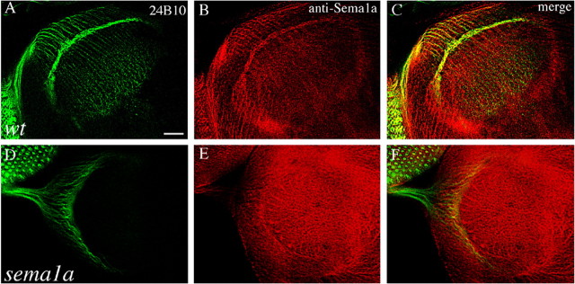
Sema1a is present in R-cell axons and growth cones in the developing visual system. A, B, Wild-type (wt) third-instar larval eye–brain complexes were double stained with MAb 24B10 (green), which recognizes all R-cell axons, and anti-Sema1a (red). The merge is shown in C. Sema1a staining is present in R-cell bodies in the eye disc and their axonal trajectories from the eye disc through the optic stalk into the developing optic lobe. The lamina plexus, consisting mainly of R1–R6 growth cones, is strongly stained. D, E, sema1aP1 mosaic eye–brain complexes in which large patches of homozygous sema1aP1 mutant cells were generated in an otherwise heterozygous or wild-type eye and were double stained with MAb 24B10 (D) and anti-Sema1a (E). The merge is shown in F. Mosaic staining pattern is observed in the eye disc and optic stalk, confirming the specificity of this antibody. Scale bar: (in A) 20 μm.

