Figure 3.
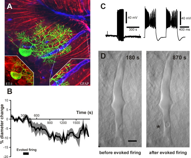
Purkinje cells express endothelin 1 and constrict microvessels. A, Confocal image of a recorded Purkinje cell (biocytin labeling; green) immunoreactive for endothelin 1 (ET-1; red; left inset) and located in the vicinity of penetrating blood vessels (immunodetected for laminin; blue). Note the processes of Bergmann glia ((immunodetected for GFAP; red) apposed to the blood vessel wall (right inset). B, Mean vascular contraction induced by 2 min of evoked Purkinje cell firing (black box; n = 6). The SEM borders the mean trace. C, Electrophysiological recording of a Purkinje cell stimulated for 2 min (left trace). An example of trains of action potentials evoked during the stimulation paradigm of the Purkinje cell, showing complex spikes (right trace), is illustrated. D, Infrared images of an intraparenchymal cerebellar blood vessel that constricted in response to the stimulation of a single Purkinje cell. Scale bar, 10 μm. The asterisk indicates a region of high vascular reactivity.

