Figure 1.
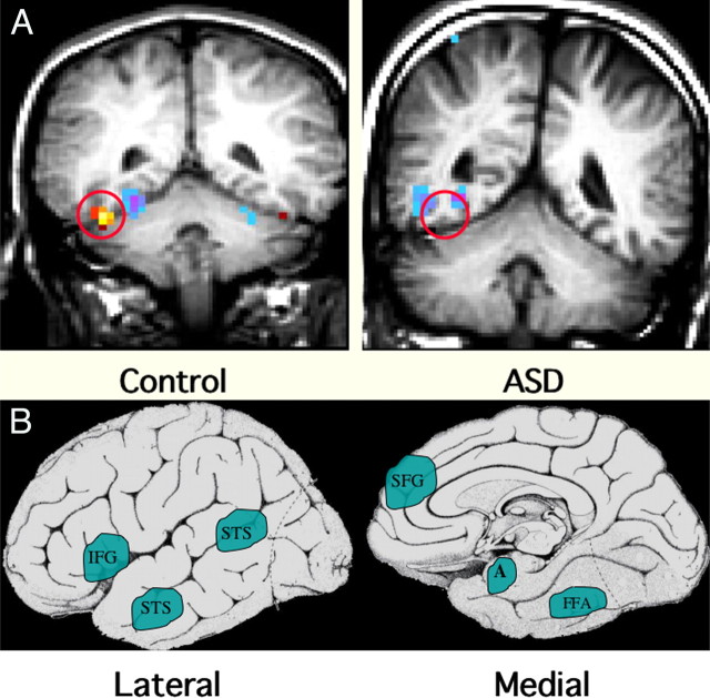
Functional MRI abnormalities observed in ASD. A, These coronal MRI images show the cerebral hemispheres above, the cerebellum below, and a circle over the fusiform gyrus of the temporal lobe. The examples illustrate the frequent finding of hypoactivation of the fusiform gyrus to faces in an adolescent male with ASD (right) compared with an age- and IQ-matched healthy control male (left). The red/yellow signal shows brain areas that are significantly more active during perception of faces; signals in blue show areas more active during perception of nonface objects. Note the lack of face activation in the boy with ASD but average levels of nonface object activation. B, Schematic diagrams of the brain from lateral and medial orientations illustrating the broader array of brain areas found to be hypoactive in ASD during a variety of cognitive and perceptual tasks that are explicitly social in nature. Some evidence suggests that these areas are linked to form a “social brain” network. IFG, Inferior frontal gyrus (hypoactive during facial expression imitation); pSTS, posterior superior temporal sulcus (hypoactive during perception of facial expressions and eye gaze tasks); SFG, superior frontal gyrus (hypoactive during theory of mind tasks, i.e., when taking another person’s perspective); A, amygdala (hypoactive during a variety of social tasks); FG, fusiform gyrus, also known as the fusiform face area (hypoactive during perception of personal identity) (Schultz, 2005; Schultz and Robins, 2005).

