
Figure 2.
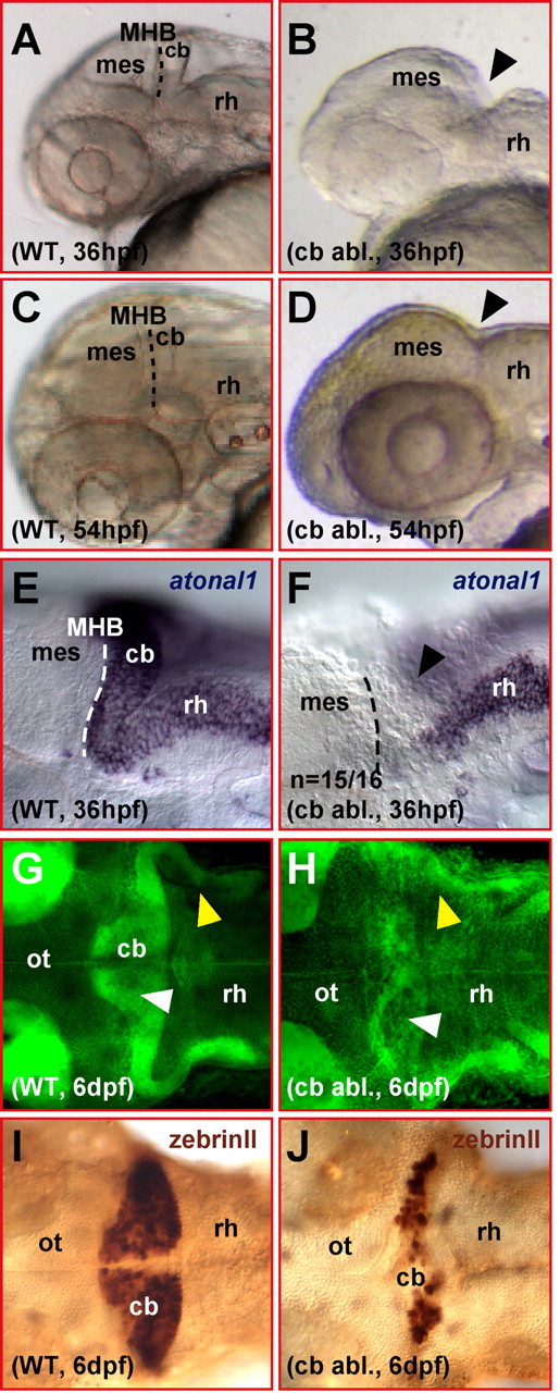
Ablated differentiating zebrafish cerebellum appears to regenerate. A–D, Lateral view of a zebrafish head showing that the mediolateral developing cerebellum posterior to the midbrain–hindbrain boundary (A, WT) is fully ablated by survival surgery (B, arrowhead) at 36 hpf. This surgical ablation leads to the fusion of the midbrain to the anterior rhombencephalon lacking a cerebellar compartment at the MHB (compare C, D, arrowhead). E, F, Lateral views. Analysis of atonal1 expression by mRNA in situ hybridization right after surgical ablation of the cerebellar tissue shows that no atonal1-expressing cerebellar cells are left behind posterior to the midbrain (F, arrowhead). G, H, Dorsal views. Bodipy Ceramide staining of cellular mem branes and neuropil reveals that larvae, which have undergone cerebellar excision (H), have formed a cerebellar compartment (white arrowhead) and cerebellar axon tracts (yellow arrowhead) at 6 dpf. I, J, Dorsal views. Furthermore, zebrinII antibody stainings reveal the presence of likely regenerated Purkinje neurons after ablation of the cerebellum at 36 hpf. cb, Cerebellum; mes, mesencephalon; ot, optic tectum; rh, rhombencephalon; WT, wild type.