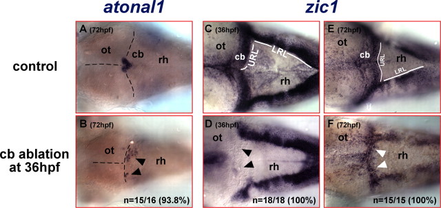
Figure 4.
The anterior hindbrain reexpresses early rhombic lip marker genes after ablation of the differentiating cerebellum. A–F, Dorsal views of the anterior rhombencephalon of embryos that have been analyzed by mRNA in situ hybridization. A, In 72 hpf wild-type embryos, atonal1 is expressed in the forming valvula cerebelli at the dorsal midline abutting the posterior optic tectum, whereas its expression in the lateral halves of the cerebellum has ceased (compare with Fig. 2E). B, In contrast, excision of the cerebellum at 36 hpf results in ectopic atonal1-expresssing cells in the lateral halves of the dorso-anterior rhombencephalon at 72 hpf. C, The zebrafish zic1 homolog is expressed in the upper and lower rhombic lips and their derived cells at 36 hpf; thus zic1-expressing cells cover the entire dorsal aspect of the cerebellum. D, Excision of the differentiating cerebellum at 36 hpf disrupts the continuous zic1 expression domain between lower and upper rhombic lips, because no zic1-expressing cells in the dorso-anterior rhombencephalon posterior to the optic tectum can be detected, indicating that cerebellar ablation leaves no URL cells behind. Strikingly, a mediolateral zic1 expression domain reappears along the new MHB in the anterior rhombencephalon after recovery at 72 hpf (F, white arrowheads) again connecting both lower rhombic lips as in wild-type embryos (E). This reexpression of atonal1 and zic1 suggests the reestablishment of rhombic lip fate in cells of the anterior hindbrain after cerebellar ablation. cb, Cerebellum; LRL, lower rhombic lip; ot, optic tectum; rh, rhombencephalon.

