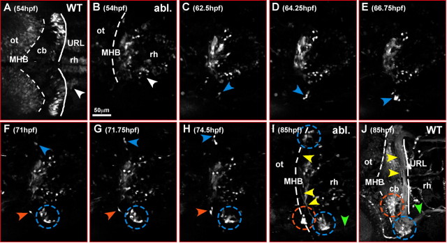
Figure 5.
Regenerating neuronal cerebellar cells are derived from the lateral anterior rhombencephalon. A–J, Dorsal views of the anterior rhombencephalon of a time-lapse microscopy recording of a transgenic 781 strain embryo, in which cerebellar excision has been performed at 36 hpf. Only embryos with complete bilateral cerebellar ablation scored through the absence of GFP-expressing rhombic lip cells on both sides of the anterior hindbrain at 54 hpf were used for time-lapse analysis. Pictures recorded at individual time points are being displayed to show the dynamics of the cerebellar neuronal regeneration processes (supplemental movie 3, available at www.jneurosci.org as supplemental material). B, At 54 hpf, GFP-expressing URL-derived neuronal precursors are absent on both sides of the anterior hindbrain compared with wild-type embryos (A), indicating that the differentiating cerebellum with the URL had been removed completely by surgical ablation. Only GFP-expressing neurons localized in the rhombencephalon posterior to the cerebellum can be detected in ventral positions (white arrowhead). C, Around 62 hpf, first GFP-expressing cells appear in ectopic locations on the far left side of the lateral dorso-anterior rhombencephalon (blue arrowhead); subsequently, GFP expression increases in these cells (D) and they migrate anteriorly toward the new MHB (E). Here they settle in a lateral position along the MHB forming a cluster of neurons (F–H, blue dashed circle) and projecting commissural axons across the rhombencephalon (I, yellow arrowheads) reminiscent of the developmental course of URL-derived neuronal cells in nonoperated transgenic gata1:GFP 781 strain embryos (J) (compare with supplemental movie 2, available at www.jneurosci.org as supplemental material). Starting ∼71 hpf, a similar regenerative event can be observed on the far right side of the anterior rhombencephalon (F–H, blue arrowhead; I, blue dashed circle). In addition, a second cluster forms through migration of GFP-expressing neurons closer to the dorsal midline (I, orange dashed circle) as in nonoperated transgenic gata1:GFP 781 strain embryos (J); similarly, axons are being projected into posterior regions of the rhombencephalon (green arrowhead). Note that there is no contribution of regenerating cells to the contralateral cerebellar one-half. cb, Cerebellum; ot, optic tectum; rh, rhombencephalon.

